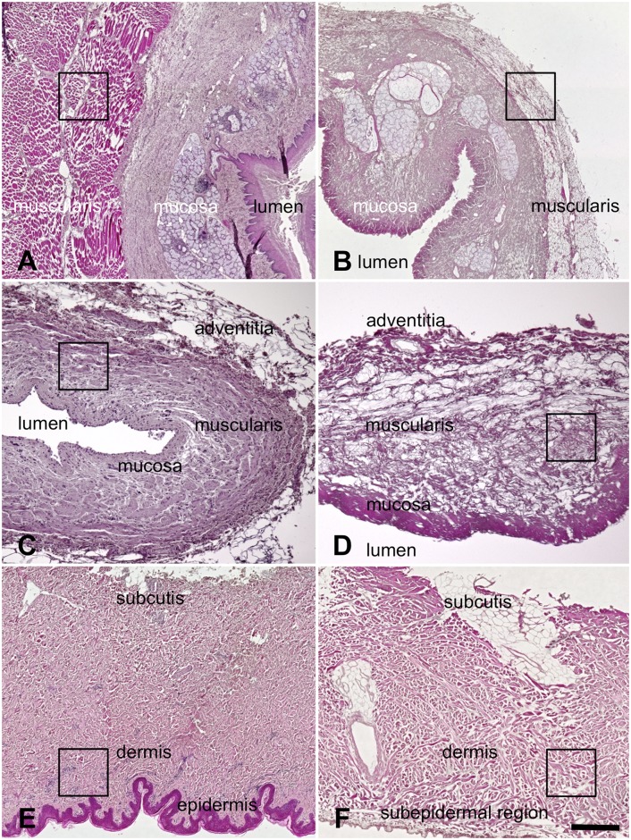
Fig 5. Hematoxylin-eosin stained-samples of 5A) native esophagus 5B) acellular esophagus 5C) native ureter 5D) acellular ureter 5E) native skin 5F) acellular skin.
In spite of the removal of the cellular structures the respective tissue layers remained intact structurally. A marked thinning was observed in the tunica media of the esophagi and some thinning in the ureters. In the acellular skin samples the epidermis was completely removed. Black rectangles indicate the regions where samples for electron microscopy were obtained (also refer to Figs 9 to 13). Scale bar: 300 μm (5A,B), 100 μm (5C,D), 300 μm (5E,F).

