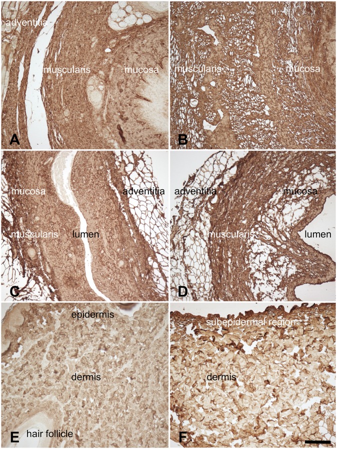
Fig 6. Anti-type I collagen staining of 6A) native esophagus 6B) acellular esophagus 6C) native ureter 6D) acellular ureter 6E) native skin 6F) acellular skin.
Collagens were observed throughout the native and acellular scaffolds but appeared to be more condensed in the latter, especially in the tunica muscularis (6B,D) and in the subepidermal regions (6F). Scale bar 100 μm.

