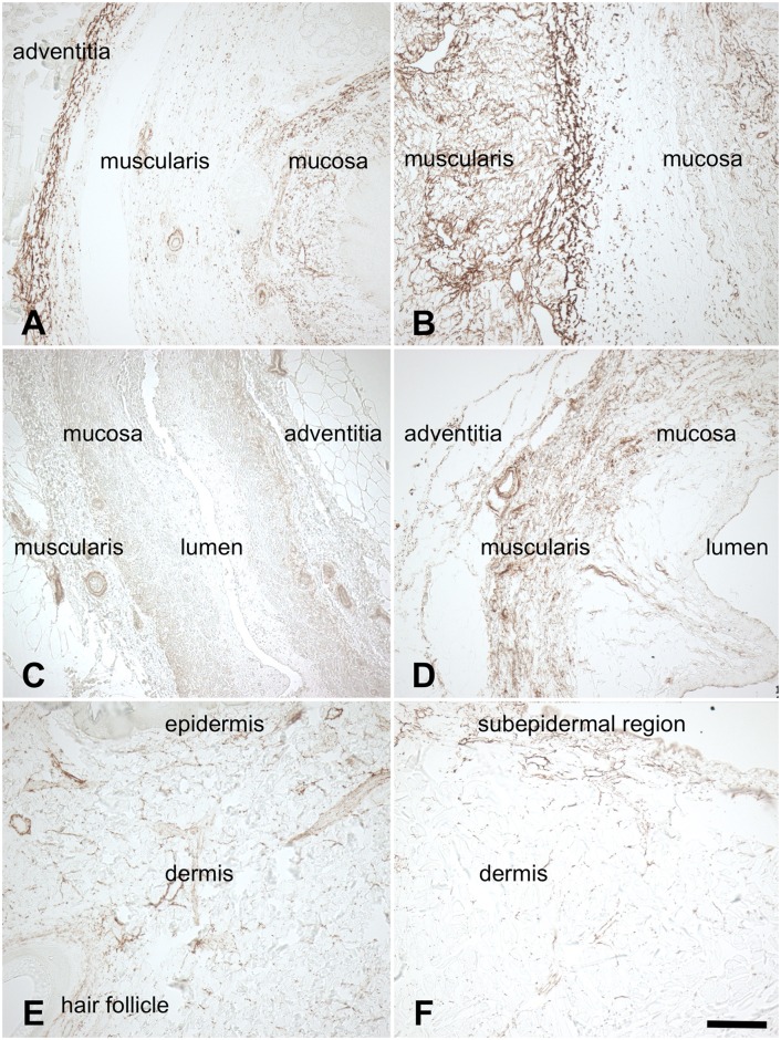
Fig 7. Anti-elastin staining of 7A) native esophagus 7B) acellular esophagus 7C) native ureter 7D) acellular ureter 7E) native skin 7F) acellular skin.
Elastic fibers were observed in the submucosal regions, the tunica muscularis, to some extent in the adventitia and in blood vessels (7A-D). The skin samples showed low quantities of elastic fibers, mostly in the vessels and dermal perivascular regions (Fig 7E,F). Scale bar 100 μm.

