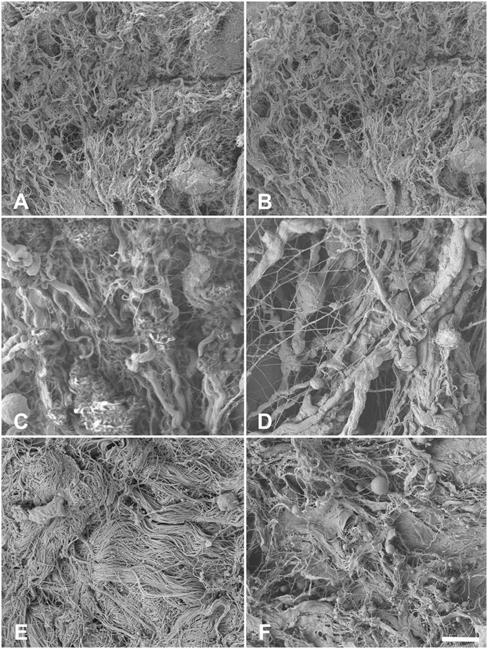
Fig 9. Scanning electron microscopy of 9A) native esophagus 9B) acellular esophagus 9C) native ureter 9D) acellular ureter 9E) native skin 9F) acellular skin.
Type I collagens appeared remain largely intact in the 1000x magnification without noticeable major direction in the tunica muscularis of the esophagi and the ureters but in the dermal skin layer. Scale bar 10 μm.

