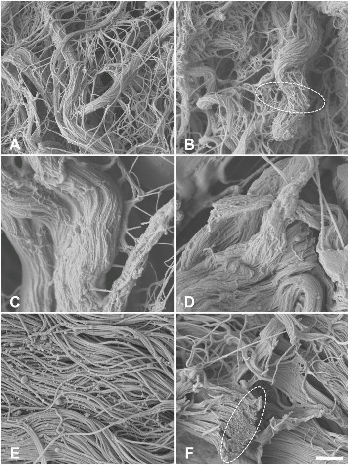
Fig 10. Scanning electron microscopy of 10A) native esophagus 10B) acellular esophagus 10C) native ureter 10D) acellular ureter 10E) native skin 10F) acellular skin.
In the higher 10,000x magnification type I collagen clotting was observed (interrupted circles) accompanied by a loss of cross-linking collagens. Scale bar 1 μm.

