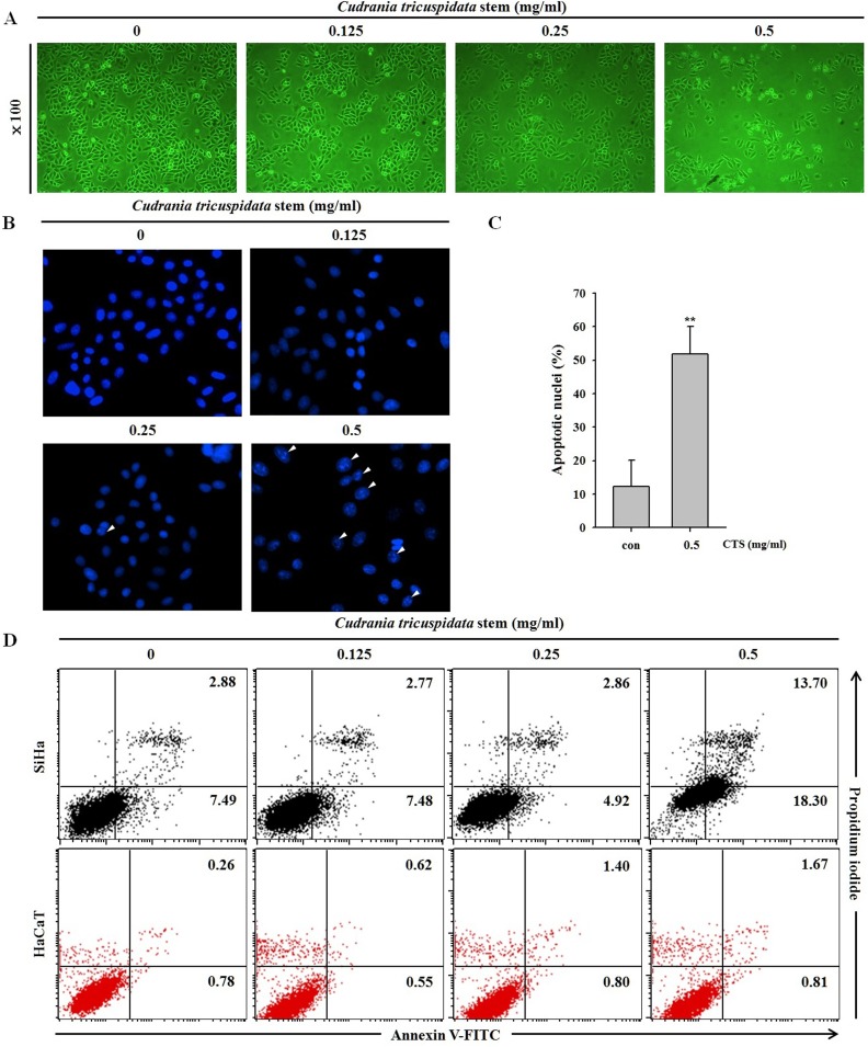
Fig 3. Effects of CTS on SiHa cervical cancer cell morphological changes and apoptosis.
(A) Microscopic images of SiHa cells treated with CTS for 24 h. The photographs were taken by phase-contrast microscopy at 100× magnification. (B) Fluorescence microscopic images of SiHa cells treated with CTS for 24 h. Nuclear condensation and chromatin shrinkage were observed. (C) The data on apoptotic nuclei of whole DAPI stained cells were summarized as bar graphs. Results of *p < 0.05 and **p < 0.005 were considered statistically significant. (D) After treatment with the indicated concentration of CTS for 24 h, SiHa and HaCaT cells were stained with annexin V-FITC/PI.

