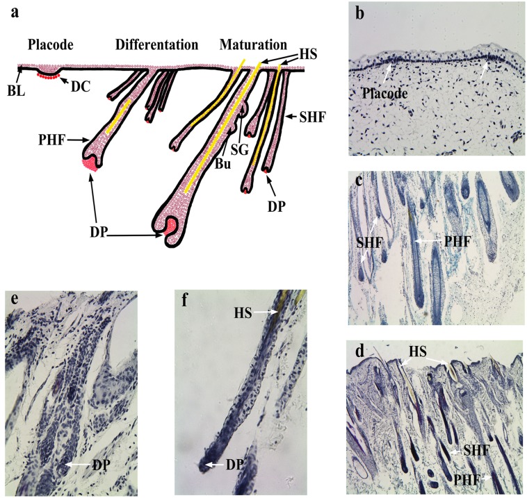
Fig 1. SACPIC staining of fetal goat tissue.
(a) Schematic representation of the process of HF morphogenesis. Longitudinal sections (× 40) of fetal HF at (b) placode (E60), (c) differentiation stage (E120), and (d) maturation stage (NB). A high magnification view (× 160) of HF at (e) E120 and (f) NB. BL: Basal layer, DC: Dermal condensate, PHF: Primary hair follicle, SHF: Secondary hair follicle, DP: Dermal papilla, Bu: Bugle, SG: Sebaceous gland, HS: Hair shaft.

