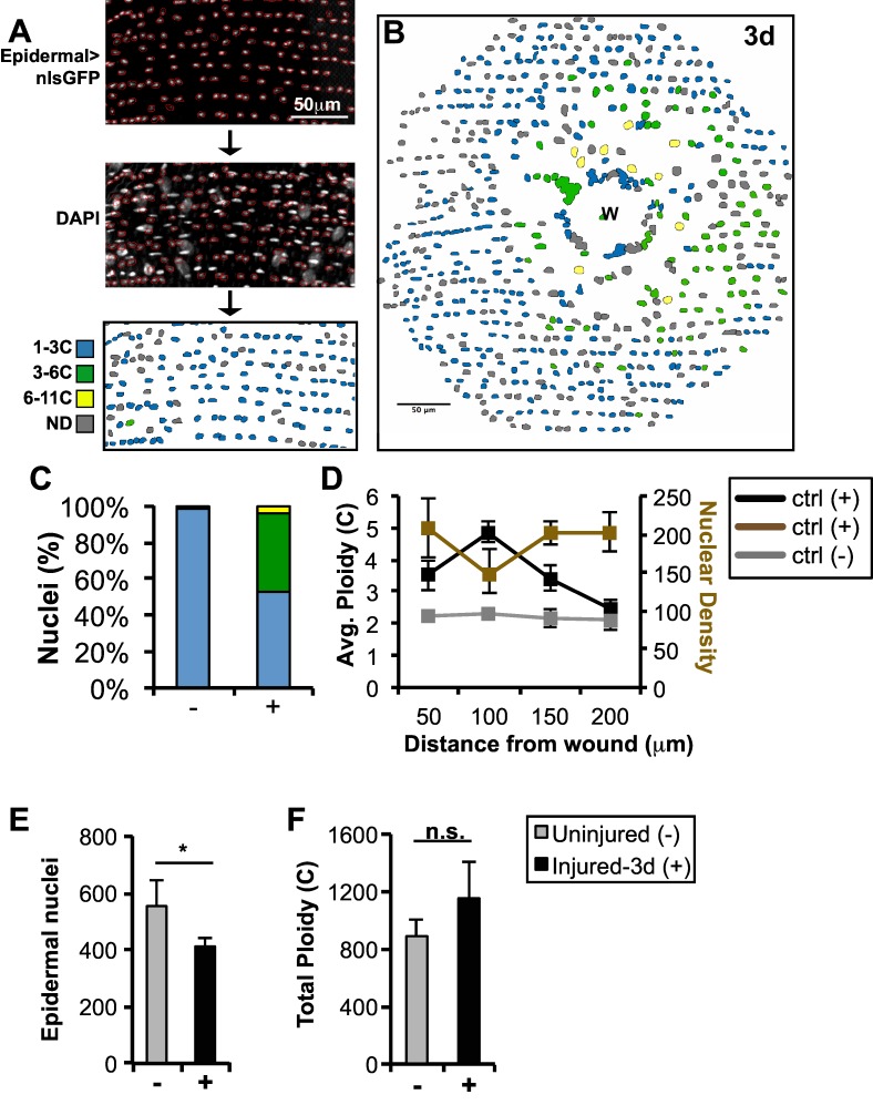
Fig 1. Wound-induced polyploidization compensates for cell loss in adult Drosophila epidermis.
Schematic of the semi-automated approach to identify and measure DNA content within fly epidermis. Fly epidermal nuclei were identified by epidermal specific expression of nuclear GFP. Nuclei were outlined (red) using Fiji’s trace function and transferred to the corresponding DAPI image. Nuclear traces were color-coded based on the normalized ploidy value as indicated. Nuclei overlapping with non-epidermal nuclei were not determined (ND, gray). Epidermal cells polyploidize in response to injury: (A) Uninjured (-), (B) 3d post injury (+), and (C) % of epidermal nuclei in the indicated color-coded ploidy ranges. (D) Average epidermal ploidy (black or gray lines for injured (+) or uninjured (-), respectively) and nuclear density (brown line) versus distance from the wound center or image center in ctrl. (E) Epidermal nuclear number is reduced 3d post injury. (F) Total epidermal ploidy is not significantly different (n.s.) at 3d post injury. All analysis was preformed with 2 uninjured and 5 injured flies. Error bars represent standard deviation where *p<0.05 is based on two-tailed Student's t test.

