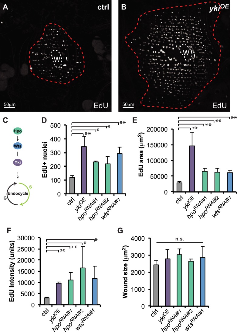
Fig 2. Hippo pathway controls the extent of endoreplication post injury.
EdU marks endoreplicating cells around the wound scar (W, white dashed line) at 2d post injury. Immunofluorescent images of EdU staining in (A) ctrl or (B) epidermal specific Yki overexpression (ykiOE). The boundary of EdU+ area is outlined (red dashed line). (C) Diagram of core Hippo signaling pathway regulating entry into the endocycle. (D-F) Quantification of the effect of core Hippo genes on EdU response where (D) represents the average number EdU+ nuclei, (E) the average area containing EdU+ cells, (F) average EdU intensity per nucleus, and (G) the wound scar size at 2d post injury. All constructs were expressed with epidermal specific-Gal4 driver and examined at 2d post injury. At least 3 flies were scored for each condition. Error bars represent standard deviation where *p<0.05, **p<0.01, and n.s., not significant (p>0.05) are based on two-tailed Student's t test.

