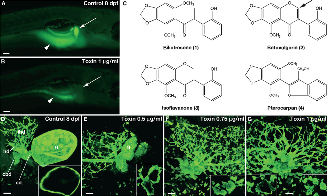
Fig. 1. Biliatresone identification.
(A and B) Lateral fluorescent image of a live 8 dpf wild-type (wt) control larva (A) and a larva treated with biliatresone (B) for 48 hours beginning at 5 dpf (1.0 µg/ml). Both larvae were soaked in Bodipy-C16 for 4 hours before imaging. Gallbladder fluorescence (arrow) is absent, and intestinal fluorescence (arrowheads) is markedly reduced in the toxin-treated larva. (C) Chemical structures of biliatresone and related isoflavones noted in the text. Arrow points to site of C-ring cleavage during possible metabolism of betavulgarin to biliatresone. (D to G) Confocal projections of the gallbladder and extrahepatic bile ducts of 8 dpf immunostained control (D) and toxin-treated (E to G) larvae. Insets show thin (1 µm) confocal sections of gallbladders. Increased doses of the toxin caused progressive changes in morphology of the gallbladder and extrahepatic ducts with preservation of the intrahepatic ducts. The variation seen in intrahepatic duct morphology is within normal limits. g, gallbladder; cd, cystic duct; cbd, common bile duct; ihd, intrahepatic bile ducts; hd, hepatic duct. Scale bars, 100 µm (A and B); 20 µm (D to G).

