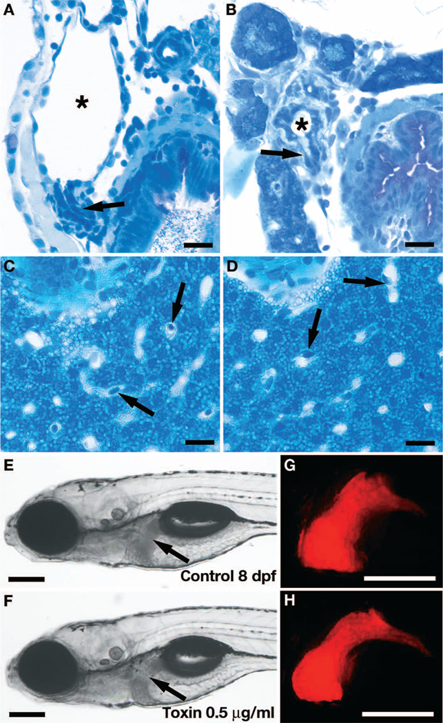
Fig. 2. Biliatresone tissue specificity.
(A and B) Histological cross sections showing severe morphological defects of the gallbladder (asterisks) and cystic duct (arrows) in an 8 dpf biliatresone–treated larva (B) compared with a control larva (A). (C and D) Normal histological appearance of hepatocytes and liver sinusoids in an 8 dpf toxin-treated larva (D) compared with a control larva (C). Sinusoids (containing nucleated red blood cells; arrows) are the principal vascular channel in the liver of zebrafish larvae; thus, branches of the portal vein and artery are not seen in these histological sections. Intrahepatic bile ducts are too small to be seen. (E to H) Bright-field images of live 8 dpf control (E) and toxin-treated (F) Tg(lfabp-RFP) larvae with corresponding fluorescent images of the liver (G and H). Arrows, liver (E and F). Scale bars, 10 µm (A to D); 200 µm (E to H).

