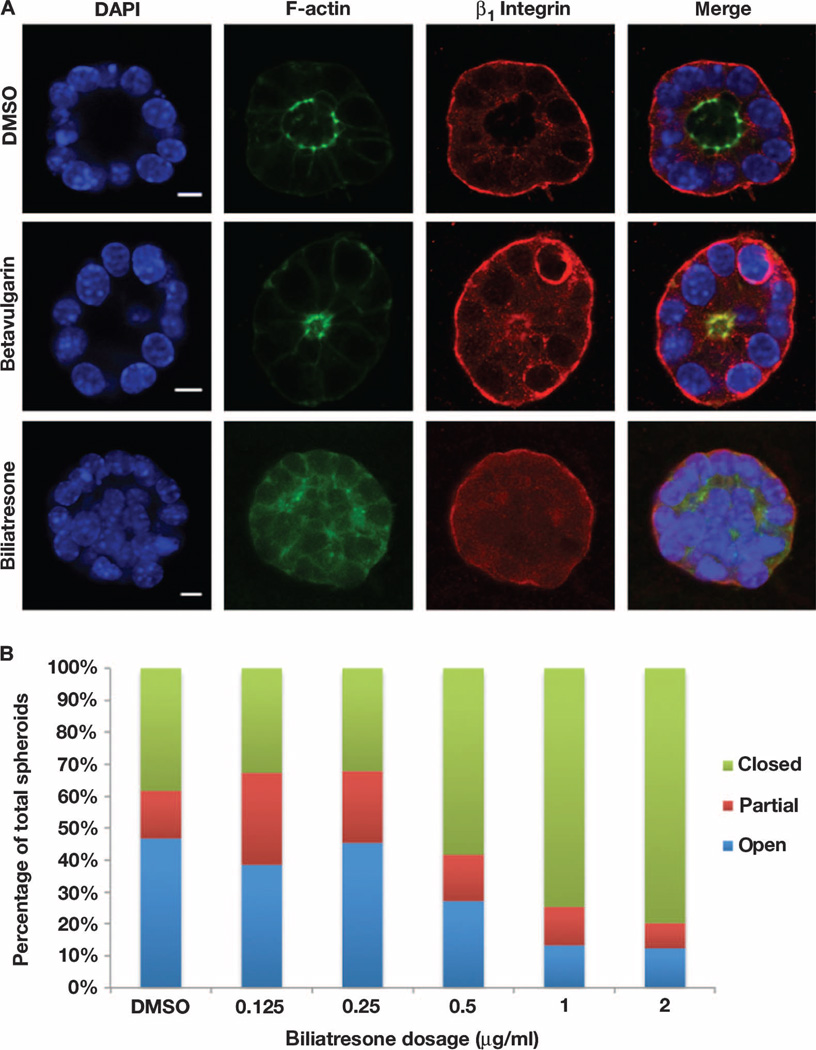
Fig. 5. Biliatresone-induced mouse cholangiocyte injury.
(A) Confocal sections through cholangiocyte spheroids treated with vehicle, betavulgarin (2 µg/ml), or biliatresone (2 µg/ml) for 24 hours and stained with antibodies against F-actin or the β1-integrin subunit, or with DAPI. The biliatresone-treated spheroid shows disruption of the spheroid lumen and abnormal cholangiocyte polarity, as evidenced by altered distribution of F-actin and by cholangiocyte stratification. Most betavulgarin-treated spheroids demonstrated decreased lumen size, but there was no loss of polarity or disruption of the monolayer (see fig. S17). Representative images from five independent experiments are shown. DAPI, 4′,6-diamidino-2-phenylindole. (B) Dose-response experiment showing progressive increase in the percentage of abnormal biliatresone-treated spheroids beginning at 0.5 µg/ml. Scale bars, 7 µm. n = 48 to 89 spheroids counted per condition.

