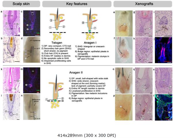
Figure 2. Telogen and anagen I, II.
For each stage, a schematic drawing is provided, and key and auxiliary features are numbered and marked.
(a–d) Telogen. HFs with typical telogen morphology represent 1–2% of all HFs on in situ findings and are generally lacking in xenografts. Key features at this stage are a very small DP and a short secondary hair germ that lacks apoptotic cells. The entire length of the telogen HF rests in the dermis.
(e–j) Anagen I. HFs with anagen I morphology are generally not found in situ but are common in xenografts, peaking on post-grafting day 33. Key features at this stage are a small DP, a secondary hair germ shaped as triangle or small crescent, and an initiation of proliferation at the base of the germ.
(k–t) Anagen II in HFs-IS (k-n) and HFs-XG (o-t). Key features at this stage are a small DP with a wide stalk (compared to telogen and anagen I), an enlarged secondary hair germ with prominent crescent shape, and a localized proliferation hotspot at the base of the germ. On in situ, the entire length of the HF rests in the dermis. In xenografts, the peak day for anagen II is post-grafting day 40.
Hosts: SCID mice (panels - f, g, h, j, o, q, r, s), nude mice (panels - e, i, p, t).
Scale bars: 100 um.

