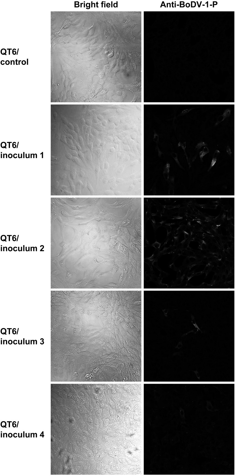
Fig. 1.
Visualization of bornaviral antigens by indirect immunofluorescence assay. Immunofluorescence assays were performed using anti-BoDV-1 P rabbit polyclonal antibodies. Bright fields and fluorescence images are shown. The cells and inocula were indicated on the left side of each panel. Inoculum 1, 12 weeks post inoculation (w. p. i.); inoculum 2, 3 w. p. i.; inoculum 3, 3 w. p. i.; and inoculum 4, 4 w. p. i.

