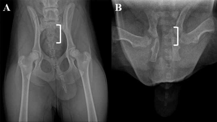
Fig. 2.

Comparison of the number of coccygeal vertebral bodies of the cloned Donggyeong dog (20 days after birth) and a donor Donggyeong dog (six months old) using digital radiographic views. A) a dorsal radiographic view of a portion of the caudal vertebral column of a cell donor dog is shown to illustrate measurements obtained for the sacrum (white bracket) through to the last coccygeal vertebra. B) dorsal radiographic view of the cloned dog. The coccygeal vertebral number was measured as the number from the dorsal surface of the sacrum. The cell donor dog had six coccygeal vertebral bodies, whereas the cloned dog had seven coccygeal vertebral bodies.
