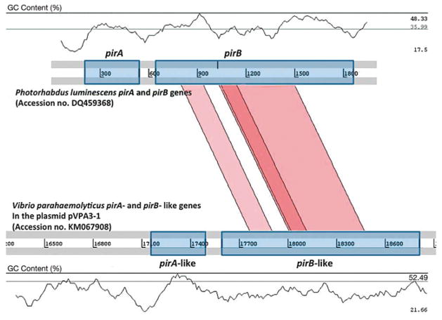
Fig. 2.
Schematic representation of the comparative analysis of the pirA- and pirB-like genes in Vibrio parahaemolyticus with the pirA and pirB genes of Photorhabdus luminescens. Regions for the pirA and pirB genes are shown with a symmetrical gene order. Translated BLAST (tblastx, score cut-off: 30) was used to align translated genome sequences. The color intensity is proportional to the sequence homology. Nucleotide base pairs are indicated between grey lines and GC contents (%) are shown

