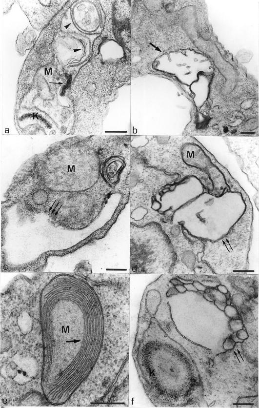
FIG.4.
(a) Thin section of a promastigote form treated with 0.1 μM compound 1b for 24 h, showing an intense mitochondrial swelling where the matrix became less electron dense. Some concentric membranes (arrowheads) and a myelin-like figure (arrows) in the matrix are observed. (b) Promastigote form treated with 0.1 μM compound 1a for 48 h, showing an intense mitochondrial swelling (arrow). (c) L. amazonensis promastigotes treated with 0.1 μM compound 1a for 48 h, showing an intense mitochondrial swelling and an expansion of the flagellar pocket (arrows). (d) Thin section of a promastigote form treated with 1.0 μM compound 1a for 24 h, showing rupture of the inner mitochondrial membrane (arrows). (e and f) Promastigote forms treated with 1.0 μM compound 1c for 48 h. These images show alterations in the mitochondrion-kinetoplast complex such as an increase in the number of mitochondrial cristae (e, arrows) and a disorganization of the kinetoplast DNA structure (f). Alterations in membranous structures, probably the endoplasmic reticulum, could be observed (f, arrows). K, kinetoplast; M, mitochondrion. Bars, 0.25 μm.

