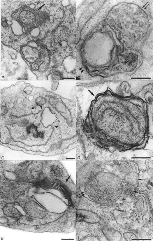
FIG. 5.
L. amazonensis promastigotes treated with 0.5 μM compound 1b and 1.0 μM compound 1c for 48 h, respectively, showing the presence of multivesicular bodies (arrows) surrounding by the endoplasmic reticulum (b, arrowheads). (c) Thin section of a promastigote form treated with 1.0 μM compound 1a for 24 h, showing rupture of the endoplasmic reticulum membrane (arrowheads). (d and e) Promastigote forms treated with 0.1 μM compound 1a and 1.0 μM compound 1c, respectively, for 48 h. These images show the presence of large myelin-like figures in the cytoplasm surrounded by the endoplasmic reticulum (arrows). (e) Structure that may correspond to the initial phase of the formation of an autophagosome, with profiles of the endoplasmic reticulum involving part of the cytoplasm (arrow). (f) Promastigote form treated with 1.0 μM compound 1c for 48 h, showing some membrane structures surrounded by the endoplasmic reticulum and containing part of the cytoplasm (arrows). Bars, 0.25 μm.

