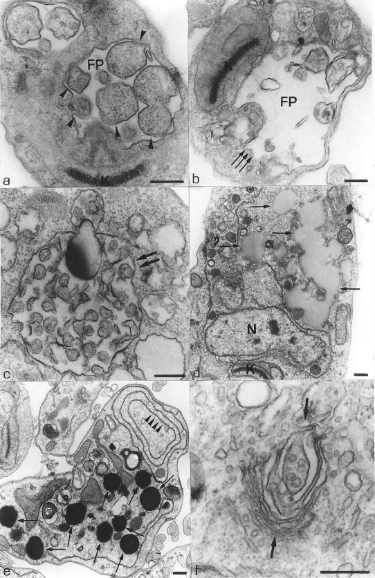
FIG. 6.
(a and b) L. amazonensis promastigotes treated with 1.0 μM and 0.1 μM compound 1a, respectively, for 48 h. These images show the presence of large vesicles in the flagellar pocket (a, arrowheads) and an intense dilatation of the flagellar pocket (b, arrows). (c) Parasite treated with 0.1 μM compound 1c for 48 h, showing the presence of a large vacuole containing some membrane profiles (arrows). (d and e) Thin sections of promastigote forms treated with 1.0 μM compound 1b for 72 h and 96 h, respectively. These images show an intense accumulation of lipid inclusions in the cytoplasm (arrows). The presence of profiles of the endoplasmic reticulum forming true vacuoles (arrowheads) is observed (e). (f) Electron micrograph of a promastigote form treated with 1.0 μM compound 1a for 24 h, showing an intense dilatation of the cisternae of the Golgi complex (arrows). FP, flagellar pocket; K, kinetoplast; N, nucleus. Bars, 0.25 μm.

