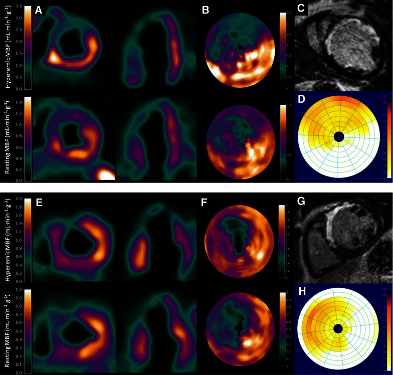
Figure 4.
Examples of [15O]H2O PET and LGE-CMR scan in two patients with a history of an anterior wall myocardial infarction. Both patients displayed significant scar size (C, D, G, and H) and perfusion defect (A, B, E and F) in the anteroseptal wall. However, quantitative MBF results showed significantly impaired hyperemic MBF in patient 2 as compared with patient 1. Note the difference in scaling on the PET images (A, B, E, and F) between both patients. Electrophysiological study showed no inducible ventricular arrhythmias in patient 1 (A-D) whereas in patient 2 (E-H) a monomorphic ventricular tachycardia was induced. CMR, cardiovascular magnetic resonance; LGE, late gadolinium enhancement; MBF, myocardial blood flow; PET, positron emission tomography. Reprint with permission28

