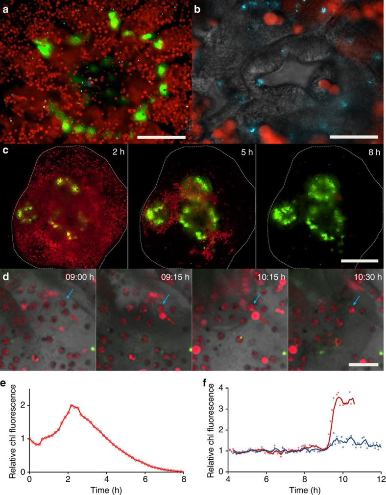
Figure 4. Using the coral-on-a-chip platform for the visualization of bacterial infection and coral bleaching.
A detailed view of microbial processes and interactions within the coral holobiont. (a) The aboral view provided by the coral-on-a-chip platform enables the visualization of fluorescently tagged bacterial cells inside the coral's gastrovascular cavity. Zooxanthellae (red) and DsRed-tagged bacterial pathogens (blue dots) are seen on the background of the coral's native GFP (green). (b) Higher magnification view of the mouth area. Algal cells and bacterial pathogens are seen on the background of the polyp's mouth, here visualized by differential interference contrast microscopy. (c) Bleaching is induced in a micropropagate subjected to high light intensity (2,500 μmol photons per m2 per s), seen through the gradual decrease in the autofluorescence signal from the algal chlorophyll. Individual polyps are seen by their native GFP fluorescence, concentrated around the mouth and tentacles. (d) Part of a bleaching process similar to a but under reduced light intensity (1,500 μmol photons per m2 per s). Algal chlorophyll and native GFP are overlaid on a bright field image of the coral tissue. This image sequence compares two individual algal cells that appear to be physically linked as one (indicated by red arrow) is lost while the other (blue arrow) remains. (e,f) Changes as a function of time of total chlorophyll (chl) fluorescence of whole micropropagate (c) and algal cells pointed by arrows (d). Values in e denote normalized chlorophyll fluorescence for the area marked by a white dashed line, corresponding to the settled micropropagate in c. Values in f denote total chlorophyll fluorescence per marked alga in d, divided by the average fluorescence for that alga from 4 to 8 h of the experiment. Red and blue curves denote algal cells pointed by red and blue arrows, respectively. Scale bars: (a) 200 μm; (b) 50 μm; (c) 500 μm; (d) 50 μm.

