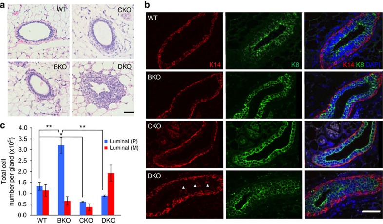
Figure 3. Altered epithelial homoeostasis in the absence of Cobra1 and Brca1.
(a) H&E staining of mammary ducts from 8-week WT and KO animals. Scale bar, 50 μm. (b) Immunofluorescence staining with luminal epithelial and myoepithelial markers K8 and K14, respectively, from 8-week animals. Scale bar, 50 μm. (c) Enumeration of mature luminal (CD49fmedEpCAMhighCD49b−) and progenitor cells (CD49fmedEpCAMhighCD49b+). The numbers of animals used are: WT (4), BKO (3), CKO (3) and DKO (4). *P<0.05, **P<0.01 by Student's t-test. Error bars represent s.e.m.

