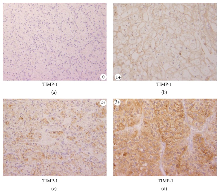
Figure 1.
0 indicates the absence of immune staining or faint membranous staining of rare tumor cells; 1+ indicates membranous staining in most tumor cells; 2+ indicates diffuse membranous and/or cytoplasmic staining in groups of tumor cells; and 3+ indicates significant cytoplasmic staining in most tumor cells. For the evaluation of immune histochemical staining, intensities of 2+ and 3+ were considered strong expressions of each protein.

