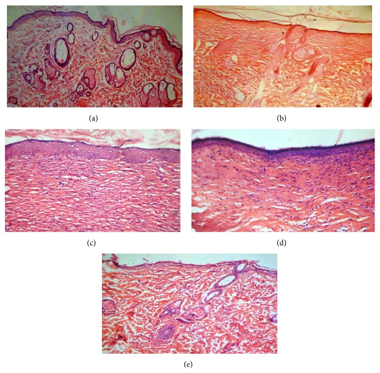
Figure 2.
Photomicrograph of histopathological section of wound tissue of rats (stained with H&E, 40x magnification). (a) Histopathological section of group I (control) animal wound tissue. (b) Histopathological section of group II (ointment base treated) animal wound tissue. (c) Histopathological section of group III (standard) animal wound tissue. (d) Histopathological section of group IV (2% w/w ointment) animal wound tissue. (e) Histopathological section of group V (5% w/w ointment) animal wound tissue.

