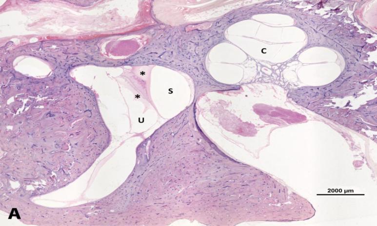
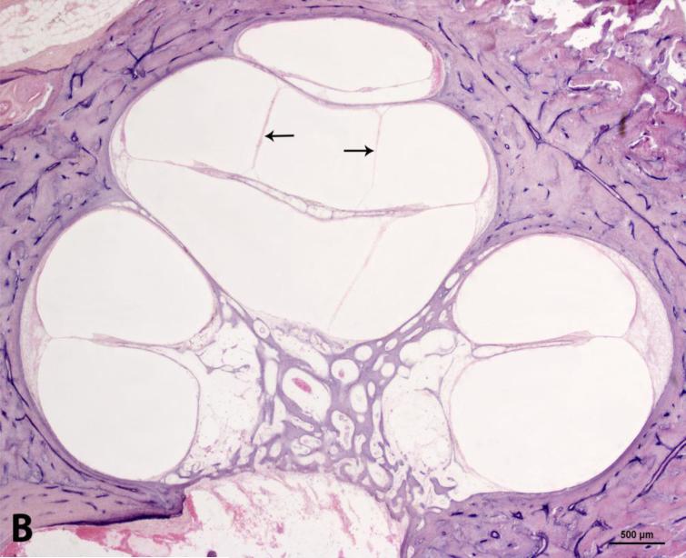
Figure 4.
Photomicrograph showing moderate endolymphatic hydrops in the temporal bone from a 71-years-old man. A. Lower magnification image shows serous labyrinthitis in vestibule. B. Higher magnification of the cochlea. C: cochlea; S: saccule; U: utricle. Arrows = endolymphatic hydrops. * serous labyrinthitis. Staining with hematoxylin-eosin (H&E).


