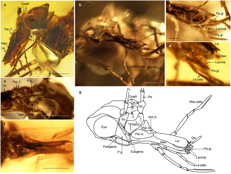
Figure 2.
Head of Psocorrhyncha burmitica gen. et sp. nov. (a) Left lateral view. (b) Dorso-frontal view. (c) Dorsal view, apex of mouthparts. (d) Lateral view, apex of mouthparts. (e) Lateral view, gena and base of mandible. (f) Dorsal view of mandibles. (g) Reconstruction of head (drawn by PN). Allotype specimen SMNS Bu-157 (a–e, g); Paratype specimen SMNS Bu-135 (f). Ant.cl. median part of anteclypeus; A.g. anterior part of gena; P.g. posterior part of gena; Ga. galea; F. frons; Fl. flagellomere; La. labrum; La. palp labial palp; Man. mandible; Max. palp maxillary palp; Pa.gl. paraglossa; Par.cl. paraclypeus; Pe. pedicel; Post.cl. postclypeus; Sc. scape, Tor. Antennal torulus. Scale bars, 200 μm (a,e,f), 100 μm (b), 50 μm (c,d).

