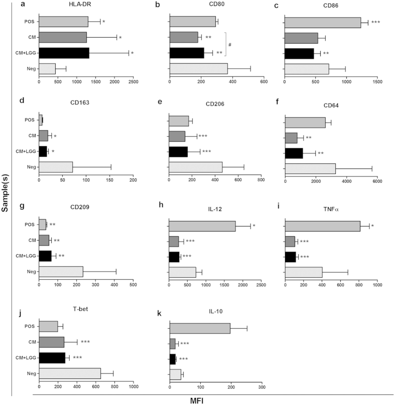
Figure 3. Expression of activation markers and intracellular cytokines in macrophages.
MFIs of activation markers and intracellular cytokines in monocyte-derived macrophages (n ≥ 4) stimulated with LGG conditioned medium (CM); or LGG conditioned medium with LGG cells (CM + LGG); or 1000 U/ml recombinant human (rh) IFNγ, 1000 ng/ml lipopolysaccharides (LPS) and 1 μg/ml R848 (positive control; Pos); or alone (negative control; Neg) were analyzed by flow cytometer after 24-hour incubation. Results were presented as mean ± SD. *p < 0.05, **p < 0.01 and ***p < 0.001 in comparisons between treated macrophages and negative control. #p < 0.05, ##p < 0.01 and ###p < 0.001 in comparisons between CM-treated and CM + LGG-treated macrophages.

