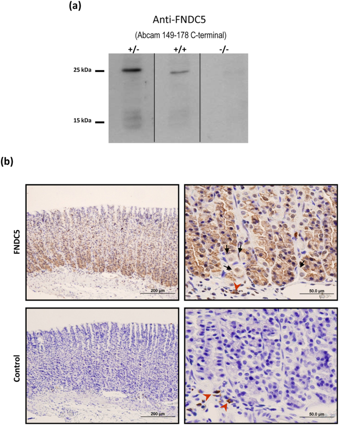
Figure 2. FNDC5 antibody specificity assay and immunohistochemical study.
(a) Blockade of the FNDC5 antibody and negative control. (+/−) FNDC5 antibody not preincubated with the immunogen, (+/+) FNDC5 antibody preincubated with FNDC5 recombinant protein, (−/−) negative control without primary antibody. Dividing lines indicate splicing of the same gel. Full-length blot are presented in Supplementary Figure 1. (b) Photomicrographs of the FNDC5 immunohistochemical studies in gastric tissue from ad libitum fed rats. FNDC5 immunoreactivity was mainly located at the bottom of the gastric glands (upper left panel). Detail of the bottom of gastric glands that show the FNDC5 positive cells (upper right panel). These cells might be identified as chief cells by their morphology and localization. The arrows indicate FNDC5 no immunoreactive cells, which were identified as parietal cells (occasional round cells with large, centrally located nuclei) (upper right panel). The corresponding negative controls are showed in the lower panels. The red arrowheads show non-specific immunoreactive cells situated at the lamina propia (probably macrophages).

