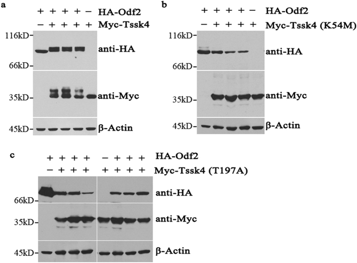
Figure 1. The association between Tssk4 and Odf2.
(a) Full-length HA-Odf2 was transfected into 293T cells either alone or in combination with Myc-Tssk4, and the electrophoretic migration rates changed for both Odf2 and Tssk4 (2nd, 3rd, and 4th lanes) when co-expressed compared with the singly transfected Odf2 (1st lane) and Tssk4 (5th lane). (b,c) Full-length HA-Odf2 was transfected into 293T cells either alone or together with two kinase-dead mutants, including (b) Myc-Tssk4 (K54M) and (c) Myc-Tssk4 (T197A). The electrophoretic migration rates of the Odf2 and Tssk4 mutants did not change. All the experiments including cell transfection, SDS-PAGE and Western blot were performed under the same experimental conditions. Since there is great molecular weight gap between HA-Odf2 (about 70kD) and Myc-Tssk4 (about 35kD), the blots are cropped to improve the clarity and conciseness of the presentation. The Western blot in all the other figures were showed in the same way.

