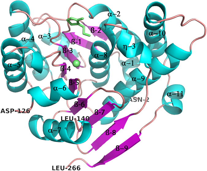
Figure 1. Overall structure of a ThiM monomer.

Cartoon representation of the SaThiM monomer. Helices are coloured in cyan and ß-sheets in magenta. The active site region with a bound THZ and a magnesium ion are highlighted and colored in green.

Cartoon representation of the SaThiM monomer. Helices are coloured in cyan and ß-sheets in magenta. The active site region with a bound THZ and a magnesium ion are highlighted and colored in green.