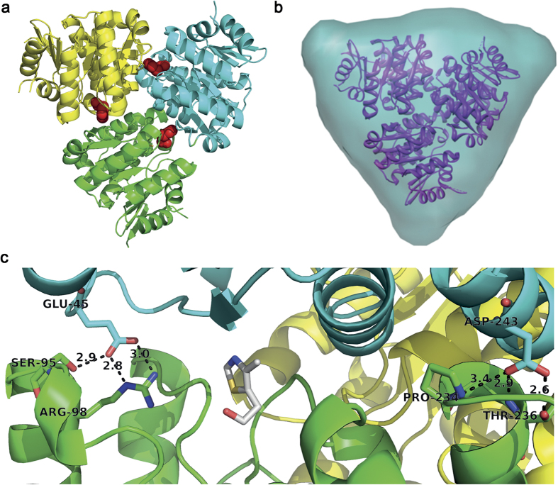
Figure 2. Structure of the ThiM trimer and its interface.
(a) Cartoon representation of the SaThiM trimer, each monomer colored differently. The three active sites are located within the interface regions of the monomers, bound THZ molecules are shown as red spheres. (b) Front view of the SaThiM trimer ab initio shape in turquoise obtained from SAXS measurements and superimposed on the crystal structure, shown in a purple ribbon representation. (c) Detailed view to the interface and active site region with functional residues shown in stick representation with carbon chain atoms in the respective chain color, nitrogen in blue and oxygen in red. THZ is also shown in stick representation with carbon atoms in grey, nitrogen in blue and oxygen in red.

