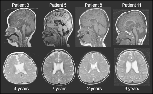
Figure 2.

Brain magnetic resonance imaging (MRI) findings of new patients with Xq28 duplications. Sagittal T1 (up) and axial T2 (bottom)-weighted images are shown. All patients showed hypoplasia of the corpus callosum and T2 signal high intensities in the deep white matter. Three patients (other than patient 8) showed atrophies of the cerebellum and bilateral dilatations of the lateral ventricles, indicating age-dependent progression. Patient 3 showed a translucent septal defect, and patients 5 and 8 showed a verga cavity.
