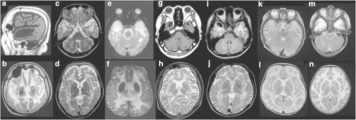
Figure 1.
Brain magnetic resonance imaging findings. (a and b) Patient 1 examined at 55 years of age. The T1-weighted sagittal image (a) shows a subcortical cyst in the anterior-temporal region, and diffuse volume loss of the cerebrum is noted (b). (c and d) Patient 2 at 51 years of age. Although subcortical cysts are too small to be detected in the anterior-temporal regions (c), mild volume loss of the cerebrum is detectable (d). (e and f) Patient 3 at 16 years of age. (g and h) and patient 4 at 30 years of age. The T1-weighted axial image (g) shows subcortical cysts in the anterior-temporal regions. (i and j) Patient 5 at 18 years of age. An asymmetric subcortical cyst in the right anterior-temporal regions is noted in the T1-weighted axial image (i). (k and l) Patient 6 at 11 months of age. (m and n) patient 7 at 9 months of age. The T2-weighted axial images indicate high intensity in the white matter in all patients (b–f, h and j–n). Subcortical cysts in the anterior-temporal regions are detectable in some of the T2-weighted axial images (e and m).

