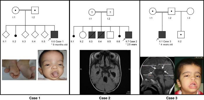
Figure 1.
Family pedigrees and clinical pictures of the three reported cases. Case 1: note the dysmorphic facial features and bilateral pre-axial lower limb polydactyl. Magnetic resonance imaging shows a classical molar tooth sign, inferior vermis hypoplasia and subsequent secondary changes in the posterior fossa and corpus callosum agenesis. Case 3: note the overlapping facial features with Case 1; magnetic resonance imaging-brain reveals dysgenesis of the corpus callosum.

