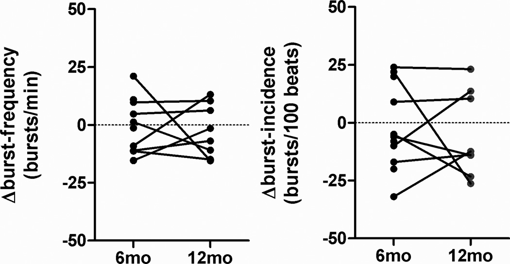
Figure 3.
Original tracings of ECG, finger blood pressure (FBP), integrated (MSNA_i) and raw multi-fiber MSNA signals. The signal to noise ratio in the raw nerve signal is sufficiently high such that single sympathetic spikes can be discriminated. Several MSNA spikes occurred outside discernible bursts in the integrated MSNA signal. Arrows mark the beginning, maximum and the end of a burst in the integrated nerve signal. Stars mark single spikes.

