
Figure 5. Comparison of the effects of tetracaine in the presence of caffeine and ryanodine .
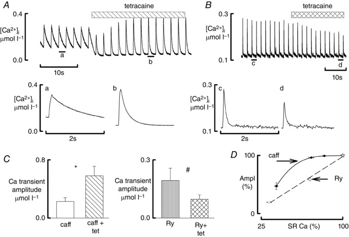
All data obtained in the presence of ISO (1 μmol l–1). A, effects of tetracaine during exposure to caffeine. Top trace shows original time course. Caffeine (1 mmol l–1) was present throughout; tetracaine (100 μmol l–1) was added as shown. The lower traces show specimen records obtained at the times indicated. B, effects of tetracaine during exposure to ryanodine. Top trace shows original time course. Ryanodine (1 μmol l–1) was present throughout and tetracaine (50 μmol l–1) was added as shown. The lower traces show specimen records obtained at the times indicated. C, mean data for the effects of tetracaine on the amplitude of the Ca transient in the presence of caffeine (left: 11 cells, 4 rats) and ryanodine (right: 7 cells, 4 rats). # P < 0.05, *P < 0.001. D, dependence of the amplitude of the systolic Ca transient on SR Ca content. Data are normalised to the control values denoted by the stars. Open symbols show the effects of ryanodine (8 cells, 6 rats) and those of caffeine (10 cells, 7 rats). The right hand ryanodine points were obtained after 3 min of exposure to 1 μmol l–1 and the left‐hand when a markedly biphasic Ca transient was seen (8–9 min). The caffeine points show (from right to left) 0.25, 0.5 and 1 mmol l–1 caffeine.