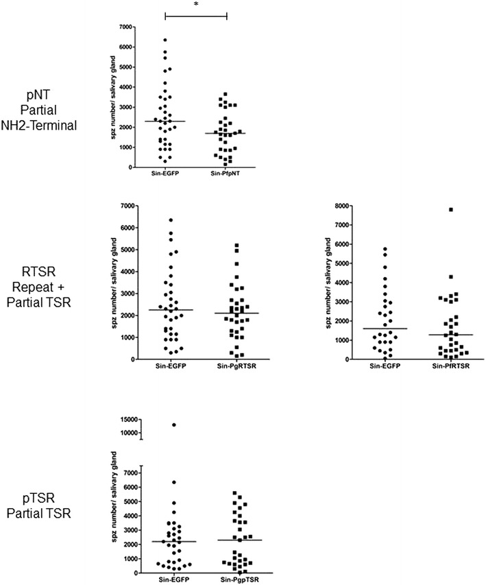
Fig. 3.

Plasmodium gallinaceum infection of individual dsSindbis-infected and control mosquitoes. Aedes aegypti females were injected intrathoracically with Sin-PfpNT, Sin-PgRTSR, Sin-PfRTSR, Sin-PfpTSR and control Sin-EGFP, and were fed three days later on P. gallinaceum-infected chickens. Salivary glands were dissected eight days after infected blood meal and sporozoite counted using phase-contrast microscopy. Solid circles (control Sin-EGFP) and squares (Sin-PfpNT, Sin-PgRTSR, Sin-PfRTSR, Sin-PfpTSR) represent individual mosquitoes with sporozoites detected in their salivary glands. The horizontal bars represent the median. A p value of <0.05 was considered statistically significant
