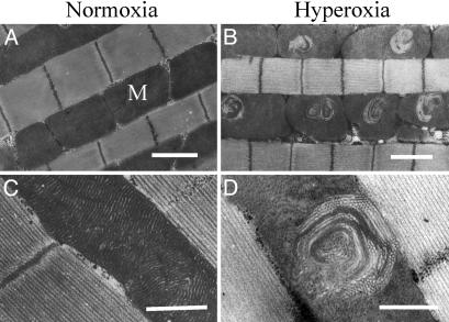
Fig. 1.
Mitochondrial swirls accumulate under hyperoxia. (A) Electron micrograph of flight muscle of Drosophila (white1118 strain) maintained for 7 days under normoxic conditions. Mitochondria (M) are aligned between the myofibrils. (B) Electron micrograph of flight muscle of Drosophila (white1118 strain) after 7 days under hyperoxia (100% O2). Mitochondrial swirls are seen in most mitochondria. (C) A normal mitochondrion at higher magnification. (D) A typical swirl at higher magnification. (Scale bars indicate 1 μm.)

