
Figure 22.
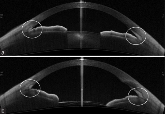
(a) Preoperative anterior segment optical coherence tomography of a case of primary angle closure glaucoma with cataract, showing narrow angles. (b) Postoperative anterior segment optical coherence tomography of the same patient following phacoemulsification showing wide open angles