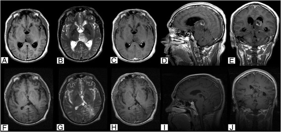
Fig. 3.

Case 1. Cranial MRI examination revealed unilateral thalamus glioma located in the left thalamus and midbrain with hypointense T1 (a) and hyperintense T2 (b) signals, which were heterogeneously enhanced after injecting contrast agent (c–e). Postoperative MRI confirmed subtotal resection (f–j). Pathological examination revealed a diagnosis of astrocytoma (WHO Grade II). Original magnification ×100
