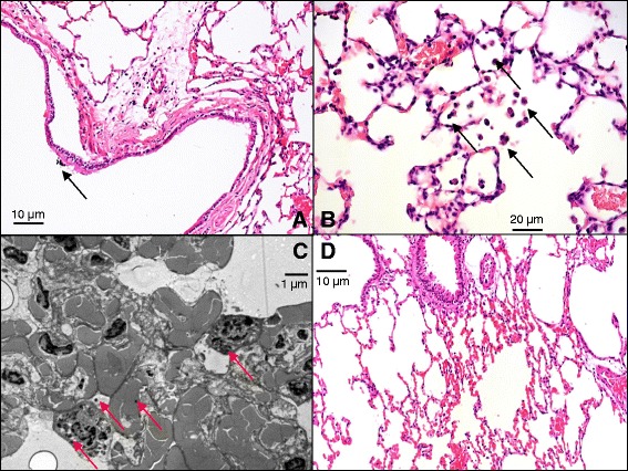
Fig. 5.

Light (a, b, d) and electron microscopic (c) images of the lungs. a: Section of a bronchus in a representative animal in the exposed group. Arrow indicates an aggregate of free dust particles inside the bronchial lumen. b: Section of the alveolar space in a representative animal in the exposed group. Arrows indicate macrophages with phagocytosed dust particles. c: Transmission electron microscopic section of a representative animal in the exposed group. Arrows indicate embedded dust particles. d: Alveolar section of a representative animal in the control group
