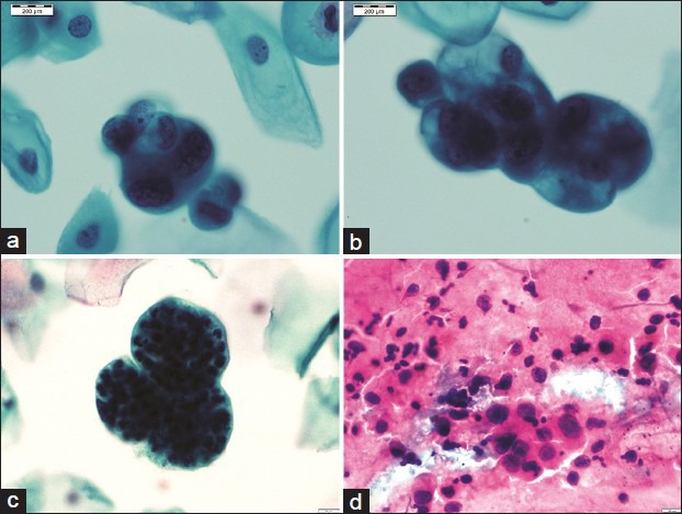
Figure 4.

A panel of microphotographs of Pap samples reported as endometrial adenocarcinoma: (a and b) Three-dimensional clusters of atypical endometrial cells showing nucleomegaly, irregular nuclear membranes, and prominent nucleoli (Pap, ×100); (c) bag of polymorphs (Pap, ×100); (d) conventional smears showing loosely cohesive cluster of atypical glandular cells with dispersed population of similar cells (Pap, ×40)
