
Figure 1.
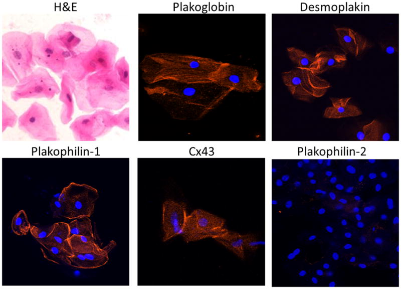
Representative images of normal buccal mucosa smears. Cells stained with hematoxylin and eosin (H&E) show typical squamous morphology with central nuclei and clearly delineated cell borders. Buccal mucosa cells immunostained with antibodies against desmosomal and gap junction proteins show strong immunoreactive signals for plakoglobin, desmoplakin, plakophilin-1 and Cx43 concentrated at the edges of the cells. No apparent signal is seen for plakophilin-2 which is expressed in the heart but not in epithelial tissues. Cell nuclei (blue) are stained with DAPI.