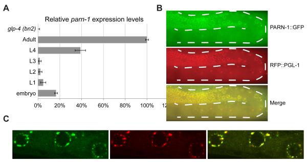
Figure 3. PARN-1 is expressed in the germline and localizes in P granules.
(A) Quantitative RT-PCR of parn-1 mRNA from total RNA isolated from synchronized wild-type populations at the indicated developmental stages and germline-deficient glp-4 (bn2) mutants at the adult stage. Expression of act-3 served as the internal control. Data were collected from three independent biological replicates. Error bars represent standard deviation.
(B) Fluorescence micrographs showing PARN-1::GFP expression (green) from an adult hermaphrodite. Expression RFP::PGL-1(red) served the P granule marker. The dashed lines outline the position of germline,
(C) Immunostaining of PRG-1 (red) and PARN-1::GFP (green) in dissected gonad arms from the parn-1 ::gfp rescue line.
See also Figure S3.

