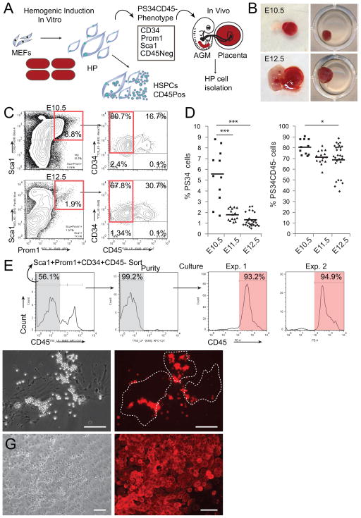
Figure 1. Prominin1, Sca1 and CD34 Mark HP Cells in Midgestation Mouse Placentas.
(A) Strategy to isolate HP cells in vivo using information generated from in vitro hemogenic induction.
(B) E10.5 and E12.5 placentas were isolated and dissociated to a single cell suspension and (C) analyzed for expression of Prom1, Sca1, CD34 and CD45, red boxes highlight populations of interest.
(D) Percentage of Prom1+Sca1+CD34+ cells (PS34, left panel) as well as the percentage of the subpopulation of PS34CD45− cells (right panel) from litters at E10.5, E11.5 and E12.5 (each mark represents a single placenta, n=11–28). *p<0.05; ***p<0.001.
(E) PS34CD45− cells were sorted and plated on gelatin-coated dishes in Myelocult with SCF, IL-3 and Flt3l for 7 days, stained for CD45 and analyzed by flow cytometry or (F) immunofluorescence. Dashed lines highlight large adherent cells associated with round non-adherent CD45+ cells.
(G) PS34CD45− cells were cultured on AFT024 stroma for 4 weeks; cobblestone areas are stained for CD45. Scale bar = 100 μm.

