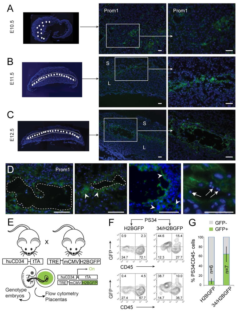
Figure 3. PS34 Cells Originate from Fetal Tissue and Localize to the Vascular Labyrinth.
Placentas from (A) E10.5, (B) E11.5 and (C) E12.5 were stained with antibodies against CD133 (Prom1, green). The left panels show composite low magnification pictures of transversal sections of placentas. The region of the labyrinth that contains Prom1+ cells is highlighted (white circles).
(D) High magnification of vasculature showing representative Prom1+ cells (arrowheads) and associated blood cells (arrows). Blood vessels are highlighted with dotted lines. Scale bar = 100 μm. S, spongiotrophoblast layer; L, labyrinth.
(E) Strategy to confirm the fetal origin of PS34 cells. Mouse placentas and embryos were isolated from crosses of huCD34-rtTA with TRE-H2BGFP transgenic mice. Embryos and placentas were isolated at E12.5. Embryos were genotyped and double-transgenic and single transgenic placentas were selected for analysis.
(F, G) Flow cytometry analysis of PS34 cells derived from double transgenic (34/H2BGFP) or single transgenic (H2BGFP) placentas. Two examples and the quantification of GFP+PS34CD45− cells in 6–7 independent placentas are shown (mean ± SD).

