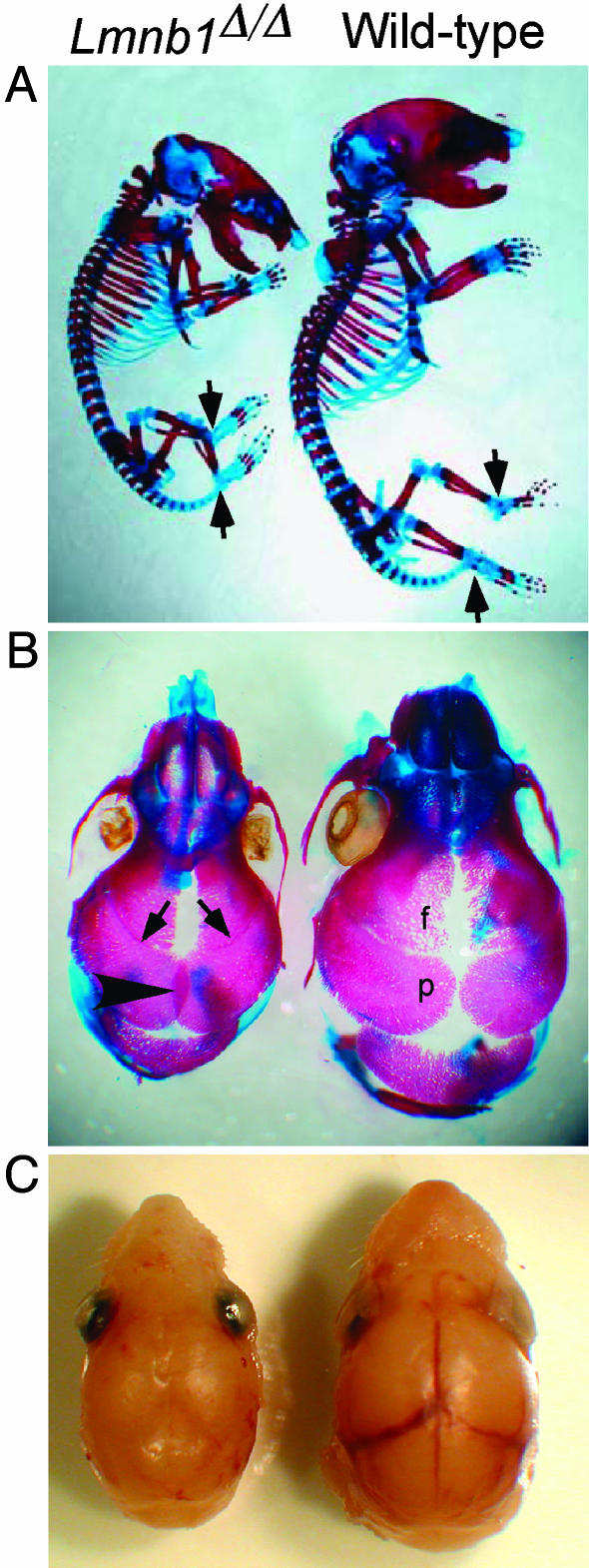
Fig. 3.
Abnormal skeleton and skull shape in Lmnb1Δ/Δ mice. (A) Alizarin red (bone) and Alcian blue (cartilage) staining of Lmnb1Δ/Δ skeleton (Left) shows a flattened skull, an abnormal vertebral column, and reduced ossification in phalanges, talus, and calcaneus (arrows). (B) Dorsal view of cranial vault. Alizarin red stain shows closure of the coronal sutures between parietal and frontal bones (arrows) and overlapping of the parietal (p) bones at the sagittal suture (arrowhead) in the Lmnb1Δ/Δ embryo (Left). (C) Removal of scalp reveals obscured view of underlying blood vessels due to suture closure in the Lmnb1Δ/Δ embryo (Left).

