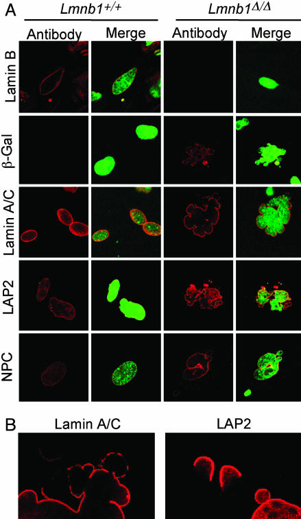
Fig. 4.
Abnormal nuclear architecture in Lmnb1Δ/Δ MEFs. (A) Confocal micrographs of MEFs from Lmnb1+/+ or Lmnb1Δ/Δ embryos stained with antibodies for nuclear proteins (Left) and merged with DNA stain for nuclei (Right). A- and B-type lamins, LAP2, and nuclear pore complex (NPC) proteins were detected with specific antibodies; the lamin B1-βgeo fusion in cells was detected with an antibody against β-gal. (B) Higher magnification of lamin A/C and LAP2 staining showing nonuniform distribution in Lmnb1Δ/Δ cells.

