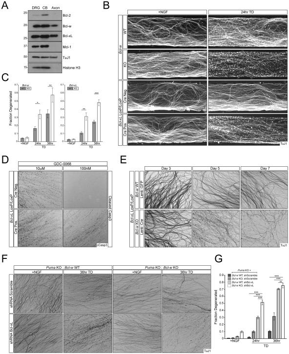
Figure 5. Bcl-xL and Bcl-w regulate axon survival.
A) Separate cell body and axon preparations were harvested from WT DRG cultures for immunoblot analysis.
B and C) DRGs from the indicated genotypes were cultured in Campenot chambers and the axonal compartment was subjected to TD for the indicated time points. Axon degeneration was visualized (B) and quantified (C). n=3 for all time points for Bcl-xLloxP/loxP. n=6 +NGF, n=5 24hr TD, and n=6 36hr TD for Bcl-xLloxP/loxP; Nestin::Cre. n=9 +NGF, n=8 24hr TD, and n=9 36hr TD for Bcl-w WT. n=9 +NGF, n=8 24hr TD, and n=9 36hr TD for Bcl-w WT. n=6 for all time points for Bcl-w KO.
D) 7 DIV Embryonic DRG cultures from Bcl-xLloxP/loxP and Bcl-xLloxP/loxP; Nestin::Cre embryos were treated with the indicated concentrations of Akt inhibitor for 12hr.
E) Dissociated and reaggregated DRG cultures from indicated genotypes were transduced with lentivirus expressing GFP or Cre. Axon degeneration was visualized at 3, 5, and 7 days post-infection.
F and G) Puma KO or Puma KO; Bcl-w KO dissociated and reaggregated DRG cultures were subjected to lentiviral-shRNA knockdown of Bcl-xL or Scrambled control. Axon degeneration was visualized (F) and quantified (G). n=2 for all Puma KO conditions and n=4 for all Puma KO; Bcl-w KO conditions.

