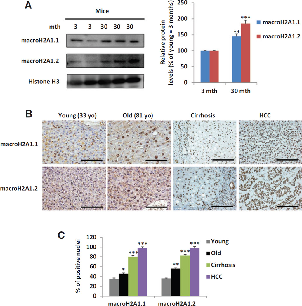
Figure 1.
macroH2A1.1 and macroH2A1.2 expression in the whole liver of: old versus young mice (A); old and young healthy controls, patients with cirrhosis and HCC (B). Age is indicated in A and B (mth, months; yo, years old).A, left, representative blots of 3 to 4 animals per age group. Histone H3 was used as a loading control; right, quantitative measurement of macroH2A1.1 and macroH2A1.2 associated signals by densitometry. Values were normalized to H3 levels and expressed as a percentage of young (3 mth). B, representative pictures of immunostaining performed for macroH2A1.1 and macroH2A1.2 in human liver samples from a young (33 years old) versus an old (81 years old) healthy subject (n = 6 per condition, 33–48 years old →young range, 72–81 years old→old range) and in human liver samples with viral cirrhosis or HCC (n = 10 per condition). All nuclei of tumor cells were positive for either macroH2A1.1 or macroH2A1.2. Positivity of hepatocytes from cirrhotic livers was intermediate. C, data are expressed as mean ± SE of 10 blindly chosen and evaluated HPF. Bar, 100 µm. A–C, *, P < 0.05; **, P < 0.01; and ***, P < 0.001.

